A Look At How Far We've Come & How Far We Have Left To Go
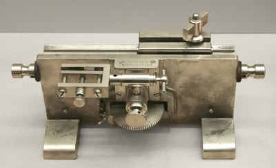
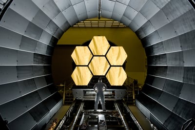
Over 150 years ago, Rudolf Jung built the first microtome for mass production in 1872, which enabled the cutting of slides for microscopic observation. At a time when people still used horses for transportation, and candles and oil lamps for lighting, this impactful invention came along and revolutionized histology and pathology. Despite its impact, the microtome has remained largely unchanged and continues to be widely used today. Fortunately, technological advancements like computers, spectral imaging, robotics, and artificial intelligence are now more readily available. Even though we possess cutting-edge tools like the James Webb Space Telescope that allow us to see deep into space, we are still examining disease tissues with visible light microscopes at 4 microns. There is still so much to discover by expanding our tools and techniques. It’s time to explore the possibilities of technological advancement and broaden our understanding of pathology beyond what can be observed by the naked eye.

est. 1600
Zacharias Janssen, a Dutch spectacle maker, is often credited with creating one of the earliest compound (two lens) microscopes.
1849
In Wetzlar, Carl Kellner establishes the "Optical Institute," marking the beginning of a remarkable journey.
1869
Ernst Leitz takes over the "Optical Institute," renaming it after himself.
1872
Rudolf Jung collaborates with Richard Thoma, bringing to life the world's first patented, mass-producible Microtome. Thus starting his precision engineering company, R. Jung in Heidelberg.

1876
Continuing his pursuit of optical excellence, Jung aligns forces with Carl Reichert, joining his optical company, C. Reichert, situated in Vienna.
1881
Horace Darwin, the son of Charles Darwin, carves his own path by establishing the optical company known as "Cambridge Instruments."
1882
Thomas Edison furthers his discovery of electricity through the invention of electricity distribution, thus illuminating the world, sparking a revolution in the way we harness and utilize this powerful force.
1886
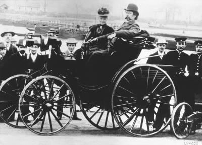
Pioneer Carl Benz introduces the world to the first-ever automobile, forever changing the course of transportation.
1921
The foundations are laid for the esteemed optical enterprise, Wild Heerbrugg, founded in Switzerland.
1953
B. Jacobson presented the initial medical application of spectral imaging (the ability to analyze tissues under a microscope without having to slice them) at the Karolinska in Switzerland.
1970s
Hyperspectral imaging, developed by NASA’s Jet Propulsion Laboratory, is refined and integrated into several scientific fields, including agriculture, environmental monitoring, food processing, and cell analysis in biology.
1972
A remarkable collaboration commences as Leitz Wetzlar and Wild Heerbrugg join hands, forging a powerful alliance that paves the way for future achievements.
1975
Expanding its horizons, Metals Research acquires Cambridge Instruments, the trailblazing pioneers of Scanning Electron Microscopes.
1986
The Wild Leitz Group is founded, forever transforming the landscape of innovation. That same year, Cambridge Instruments acquires Reichert-Jung, solidifying its position as a leader in the industry.
1990
Cambridge Instruments and The Wild Leitz Group merge together, to become the Leica group.

2004
Creation of the James Webb Space Telescope starts, manufactured by Northrop Grumman, Ball Aerospace, & L3Harris, with its completion in 2011, expanding our understanding of the universe.
2022
Celebrating the 150th anniversary since the invention of the microtome, we reflect upon the countless contributions and milestones that have shaped the realm of precision engineering, optics and the world.
As we celebrate the 150th anniversary of the microtome's invention, we are reminded of the profound impact it has had on the field of histology. The ability to prepare tissues for observation under a microscope has advanced our understanding of diseases, leading to improved diagnostics and treatment strategies. However, while technological advancements in various fields have reshaped our world, we have yet to fully explore the untapped potential beyond the microtome, and the microscope. With the accessibility of cutting-edge technologies like spectral imaging, robotics, and artificial intelligence, we stand on the precipice of unlocking new frontiers in histological research and pathology. By embracing these advancements and pushing the boundaries of what is possible, we can unravel the mysteries of pathology and propel the field into a future where precision and innovation intertwine.
Head to Barnes-Jewish Hospital's LinkedIn Post to checkout a demo of a histotechnician using a microtome to section tissue segments for microscopic observation.


